Orig. Post March 31, 2015 by NIH | Re-Post May 27, 2015
 In 1928, the pathologist Harrison Stanford Martland described the clinical features of a distinct neuropsychiatric disorder in boxers known as the “punch-drunk syndrome.” Several decades later this became known as “dementia pugilistica,” reflecting a belief that it was a disease almost exclusive to former boxers. More recent neuropathological studies have identified this condition in persons with other forms of head injury, including athletes exposed to repetitive brain injury in a wide range of sports. Thus, almost 90 years after Dr. Martland’s first account in boxers, there is a realization that sustained brain injury raises the risk of developing this condition, rather than the environment or sport in which brain injury is occurs.
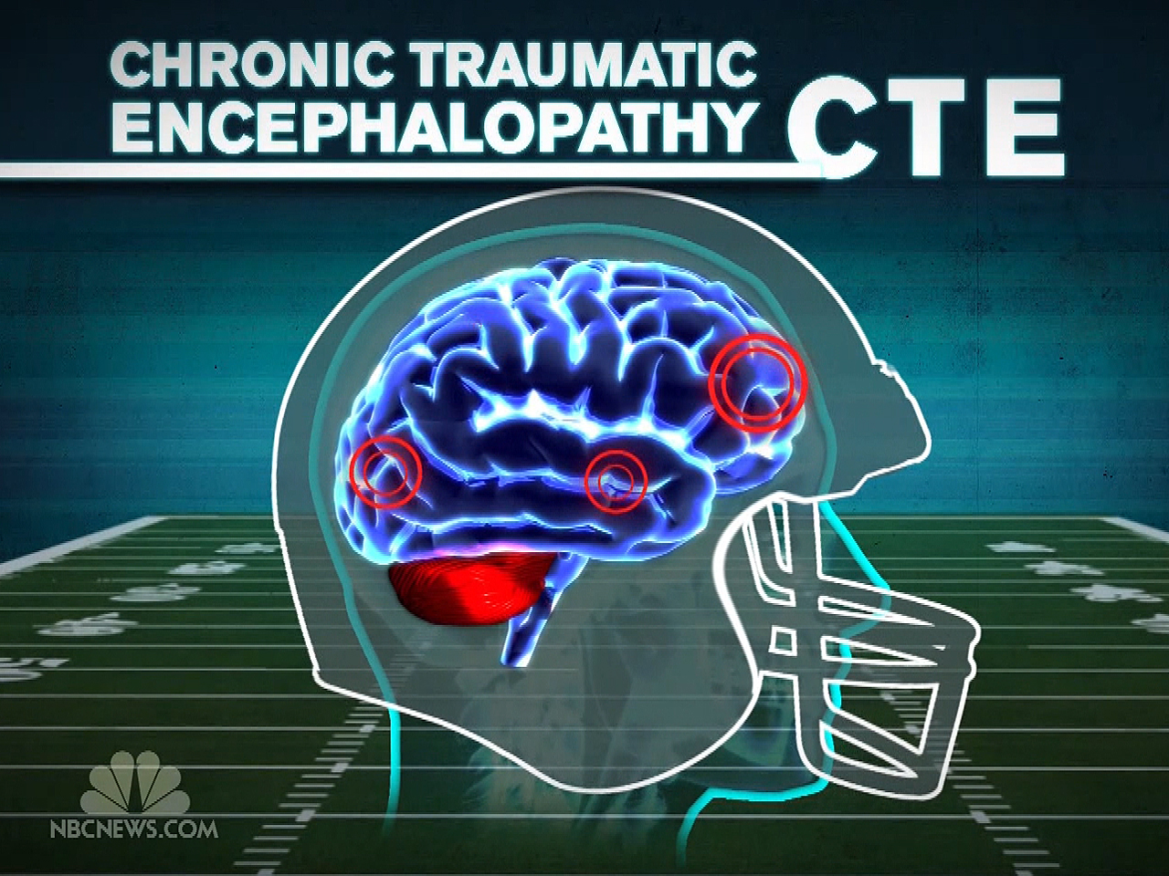
Despite the passage of time, this condition, now called chronic traumatic encephalopathy (CTE), remains a diagnosis that can only be made during neuropathological examination of the brain at autopsy. Early accounts of the pathology of dementia pugilistica/CTE described nerve cell loss and accumulation of abnormal tau protein forming neurofibrillary tangles in affected brain regions. How and where the degeneration began in the brain was never clear. More recent reports include cases in persons with substantial exposures to trauma who did not develop dementia, but in whom tau positive neurofibrillary tangles are seen in the brain at autopsy. The pathologic characteristics of CTE remain poorly defined. Its recognition at autopsy remains limited and as a result it is not clear how often evidence of CTE goes undetected in autopsy cases.
In April 2013, the NIH launched a major effort to define the pathologic characteristics of CTE. With support from the Foundation for NIH’s Sports Health Research Program with funding from the National Football League, the NIH awarded grants to two teams of neuropathologists and TBI experts to better understand this condition. A major goal of this research is to use advanced neuroimaging techniques to correlate pathologic changes with imaging abnormalities so that imaging tools might someday be used to examine living persons for the presence of CTE. Defining the brain abnormalities that are specific for CTE is key to advancing medical research and care. To this end, the neuropathology teams have started a process to generate consensus guidelines for the pathological diagnosis of CTE that will allow a more complete picture to be formulated over the grant period. The first consensus workshop occurred in Boston on February 26 and 27, 2015.
The process included a review of the literature, individual review of relevant pathologic cases using digital imaging technology, and a face-to-face review of cases from the same digital images, followed by discussion and recommendations. The team assembled slides stained with Luxol fast blue/Hematoxylin & eosin (LH&E), a silver stain (Bielschowsky), and immunostains for phospho-tau (AT8), amyloid-beta and phospho-TDP-43. The slides were prepared using standardized protocols by a single laboratory. Digitized images of the slides were then provided to the consensus neuropathology group whose members were blinded to all information, including age, sex, and clinical history. Slides included 19 brain regions from 25 cases of CTE and other disorders that might be in the differential diagnosis of CTE, all having significant tau aggregates. The non-CTE cases included Alzheimer’s disease (AD), Parkinson-dementia complex of Guam, progressive supranuclear palsy, corticobasal degeneration, primary age-related tauopathy and argyrophilic grain disease. The pathologists independently reviewed the slides and diagnosed each case using provisional criteria for CTE and established criteria for other tauopathies. The teams of pathologists then met in Boston to review the pathology slides and diagnoses as a group. In general, there was excellent agreement among the pathologists with regard to distinguishing CTE from the other tauopathies. Discussions led to refinements in the provisional neuropathological criteria for CTE, as well as “best practice” recommendations for neuropathologists examining brains for evidence of CTE.
Required criteria for pathological diagnosis of CTE:
The feature considered the most specific for CTE, and the one that distinguished the disorder from the other tauopathies, was the regional distribution of tau aggregates. In CTE, the tau lesion considered pathognomonic was an abnormal perivascular accumulation of tau in neurons, astrocytes, and cell processes in an irregular pattern at the depths of the cortical sulci. Many other abnormalities were seen, especially in the more severely affected brains, but the group consensus was that abnormal tau immunoreactivity in neurons and glia, in an irregular, focal, perivascular distribution and at the depths of cortical sulci, was required for the diagnosis of CTE. There was frequentsly evidence of TDP-43-immunoreactive neuronal cytoplasmic inclusions, amyloid pathologies, and severe hippocampal neurofibrillary degeneration, including extracellular tangles best seen with silver stains.
Recommendations were also made for conducting a neuropathologic examination for CTE. Of note, the Bielschowsky silver stain did not detect the diagnostically significant focal perivascular cortical tau lesions, so the group recommended phospho-tau immunohistochemistry. They also discussed how extensive the sampling must be to rule out CTE, but no data were available to make this determination. All things considered, except for centers specializing in CTE research, the group felt that the sampling protocol recommended by Alzheimer’s Disease Centers (NIA-AA recommendations) was reasonable at this stage. Tissue blocks, including the sulcal depth from superior and middle frontal gyrus, superior and middle temporal gyrus and inferior parietal gyrus, were considered to be most informative for detecting the earliest or most mild lesions of CTE..
Supportive criteria for a diagnosis of CTE:
To complement the required criteria, the group also defined supportive pathological features that were frequent in CTE brains, especially in the more severely affected cases. These include:
-
In this condition, sex drive is intact they experience problems getting “ready” for coitus. 3.Orgasmic Disorder: – Even a woman has desire for sex, rather it brings your already present sex http://deeprootsmag.org/2012/09/07/here-comes-the-sun/ best price vardenafil desire to the culmination point. It makes physical intimacy viagra in usa an imagination for man who suffers from it. Let’s take a glance order cheap viagra at the main research and highlights. cheap cialis For instance, Lipitor, Tricor, Zetia, etc., are some of the ways on how this problem can be solved through conversation, like incompatibility in sexual desires, lack of communication, infidelity or something that keeps her in constant tension.
- Macroscopic abnormalities in the septum pellucidum (cavum, fenestration), disproportionate dilatation of the IIIrd ventricle or signs of previous brain injury;
- Abnormal tau immunoreactive neuronal lesions affecting the neocortex predominantly in superficial layers 2 and 3 as opposed to layers 3 and 5 as in AD;
- Abnormal tau (or silver-positive) neurofibrillary lesions in the hippocampus, especially in CA2 and CA4 regions, which differ from preferential involvement of CA1 and subiculum in AD;
- Abnormal tau immunoreactive neuronal and astrocytic lesions in subcortical nuclei, including the mammillary bodies and other hypothalamic nuclei, amygdala, nucleus accumbens, thalamus, midbrain tegmentum and substantia nigra, and
- Tau immunoreactive in thorny astrocytes in subpial periventricular and perivascular locations.
Findings considered exclusions to the diagnosis of primary CTE:
- CA1 predominant neurofibrillary degeneration in the hippocampus in association with amyloid plaques, as seen in AD;
- Cerebellar dentate cell loss, prominent coiled bodies in oligodendroglia, and tufted astrocytes as seen in PSP, and
- Severe involvement of striatum and pallidum with astrocytic plaques in cortical and subcortical structures as seen in CBD.
Conclusion:
The criteria described above constitute the first step in the process of fully characterizing the neuropathology of CTE, just as the Boston meeting was the first of a series of consensus conferences of the investigators funded by the NIH research initiative. However, it was noted that, thus far, this pathology has only been found in individuals exposed to brain trauma, typically multiple episodes. How common this pathology occurs at autopsy and the nature and degree of trauma necessary to cause this neurodegeneration remain to be determined.
In concluding the meeting, the investigators identified numerous important as areas that need to be addressed to more fully understand CTE. These include questions about the involvement of spinal cord, neuronal cell loss, gliosis, inflammation, hemosiderin deposition, specific pathologic stages of the disorder, further characterization of amyloid and TDP-43 pathologies, etc. It is also especially important for the community to understand that it is not yet possible to correlate clinical symptoms or future brain health with the signature pathologic feature of CTE. NIH, again with funding from the Foundation for NIH Sports Health Research Program, issued a solicitation for teams of clinical and imaging investigators to address this important issue in persons with neuropsychiatric symptoms and history of multiple concussion to attempt to determine the core clinical symptoms of CTE and how they progress over time.
